Blog

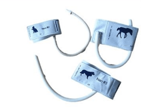
We know animals and we care of them. In May, we donated a vital signs monitor, VS2000VCS, with sidestream ETCO2, ECG, SPO2, Temperature, NIBP. We also provide 2 years free maintenance and accessories. High quality, reasonable price, excellent service are our basic policy. The VS2000VCS Veterinary vital signs monitor is a Multi-parameter monitor. It is with parameters of SPO2, ECG, NIBP, PR, RESP, TEMP, and ETCO2. It is highly compact and portable to carry out. Also it is providing fast and reliable measurements for the veterinary patients. The measuring range from the small to big animals such as cats dogs, horses. It is suitable for vet hospital, clinic, and animal labs. It has been sold to more than 30 countries such as the United States, Korea, and Colombia and so on. Both retailers and veterinarian are satisfied with our products in terms of quality, price and prompt after-sales service. Main Features: Warranty: Two years for mainunit, 6months for accessories; 7.1' coloured and bright, high definition (800 x 480) TFT LCD screen; Parameters: SpO2, ECG, Heart Rate,NIBP, TEMP*2, RESP (Optional: ETCO2) Suitable for cat, dog, horse patients ; 72 hours graphic and tabular trends of all parameters; 32 seconds full-disclosure waveform review 500 NIBP and SpO2 measurement data can be stored and recalled; 3-level audible and visual alarms and alarm events can be stored and reviewed. 100M Base-T Ethernet Interface, Support to connect to IPMS8000 Patient Management System; Special designed SpO2 technology with adjustable emitting light and a wide range measurement for veterinary Special designed ECG technology with high input impedance to suit for different size of veterinaries Special designed NIBP technology with different size of NIBP cuffs for veterinaries Sidestream technology accommodates intubated and non-intubated animals Unique Designed LOGO for veterinary use only Ergonomically designed handle to fit your hand comfortably Can link with IPMS8000 intelligent patient monitor system Multi-language: English, Chinese, French, Spanish, Germany STANDARD PACKAGE: VS2000V main unit(ECG+NIBP+SpO2+RESP+TEMP+PR) Veterinary SpO2 sensor (Y type Clip) 5-lead ECG cable 5 ECG Clips NIBP cuffs for Veterinary(Cat,Dog, Horse) NIBP Extension Tube Temp Rectal Probe Sidestream ETCO2 Sensor Nasal Sampling Line Gas Sampling Line Gas Dryer Line T-fitting Tube Sidestream Adapter Lithium Battery Power cord User Manual

Many monitors have a memory storage function. If the monitor was used to measure a hypertensive animal last time and it was not cleared to zero after use, the next time when measuring another animal with normal blood pressure by the same monitor. The blood pressure is likely to be high for the first measurement due to the automatic calibration function. It will be close to the animal's actual stae at the second time.
Intravenous infusion is a commonly used method of administration in clinical treatment. The speed of intravenous infusion varies according to the nature of the drug and the patient's physique. If the infusion is too fast or too slow, it is difficult to achieve the desired therapeutic effect and even affect the safety of patients. The infusion pump is an instrument that can accurately control the number of infusion drops or the infusion flow rate, ensure that the drug can enter the patient's body evenly, accurately and safely, and function. The speed is not affected by human blood pressure and the operator, and the infusion is accurate and reliable, which helps reduce the intensity of clinical nursing work, improve the accuracy, safety and quality of infusion. The infusion pumps have various product models and performances. According to its working characteristics, there are three types: peristaltic controlled infusion pumps, fixed volume controlled infusion pumps and syringe microinjection infusion pumps. IP300V (veterinary infusion pump) uses microelectronics technology. By accurately controlling the stepper motor and its peristaltic transmission device, the infusion pipeline is squeezed to achieve accurate control of the infusion speed and monitor the infusion process, which can greatly improve the safety and accuracy of infusion reducing the labor intensity of staff. MAIN FEATURES Delivery range: 0.1ml/h~1200ml/h. High accuracy +/- 2% after correct calibrated. 5 work modes: Rate Mode, Time Mode, Dose Mode, Sequential Mode and Drug library Mode. Manual or Automatic Bolus /Purge/KVO function can be selected. Vertical & Horizontal mounting clamp. Anti-bolus system to reduce significantly after occlusion sudden release. DPS dynamic pressure display. 5000 events stored can be checked and downloaded. Large LCD display, the brightness can be adjusted to adapt to the clinical use of the environment. IPX4 high waterproof rating for all clinical application scenarios Panel is easy to clean to ensure clinical cleanliness Infusion speed can be adjusted at any time, even the pump is running Large-scale LED colorful alarm light
The BestScan® S10, app-based WIFI color Doppler ultrasound system, comes with Colour Doppler and Colour Power Doppler as standard.The portability, tough construction and high-tech design of the BestScan® S10, it answering the demands of today and tomorrow veterinarians. Excellent image quality The BestScan® S10 provides excellent image quality in a wide range of applications. Besides B-mode, M-mode and Doppler imaging, the system offers multiple technologies to meet every clinical need. Durable and tough Unrivalled durability, extremely lightweight (0.7 Kg) design and ability to run on mains or battery power makes the BestScan® S10 easy to use anywhere, anytime. Simplified Workflow The ergonomic app-based smart device operation allows for easy manipulation of image settings, especially useful in crowded environments or inside vehicles when space is a premium. Measurements and Reports Confirming BMV’s dedication to the veterinary world, complete veterinary measurement packages, including abdominal, cardio-vascular, and reproductive gestational calculations provide easy examination documentation. TRANSDUCERS Support 5-1 MHz Phased Applications: Cardiology in dogs, cats, and exotic animals 8-5 MHz Curved Applications: Abdominal scanning and basic cardiology in dogs, cats, and exotic animals 10-5 MHz Linear MSK in dogs and cats and general imaging of exotic animals 5-2 MHz Curved General imaging in large dogs and large-sized exotics 10-5 MHz Linear For all frequently occurring cases in daily large animal practice. This probe is excellent for reproductive and basic tendon scanning 5-2 MHz Curved This probe is wide field of view and deeper penetration,excellent for daily large animal practice. Biopsy guides are available as an option for all relevant transducers I nnovative ergonomic design in every details We’ve built robust S10 to withstand dirty farm environment and other unpredictable environments, also we have a range of smart viewing options such as smart deivce and goggles, and optional accessories such as wireless charger, IFR introducer, chest mount, wrist mount, and so on. Rugged design, easy to clean Design in line with operator's habits, simplified interface Lithium battery power supply for up to 12 hours 128 elements, receiving on 32 channels, 26 frames per seconds Record videos and save images on your smart device SYSTEM SPECIFICATIONS System weight: 12.6 lbs./5.70 kg with battery Dimensions: 44.7 cm x 29.3 cm x 12.3 cm Viewing device: Smartphone / Tablet/ I-Scan® Goggles. S10 Scanner wireless link to compatible viewing device Display(Tablet): 12.1”/30.7 cm diagonal LCD Viewing Angles: 85° up/down/left/right The 2GB of tablet internal memory allows you to store approximately 2,000 images. Transfer of images to a PC is easy using wifi,bluetooth,USB. Architecture: All-digital broadband Dynamic range: Up to 165 dB Gray scale: 256 shades Application of Color Doppler Ultrasonography in Bovine and Equine Reproduction Doppler ultrasound is an emerging technology that has the potential to increase the diagnostic, monitoring, and predictive capabilities of bovine and equine theriogenologists and researchers. For example: We can use S10 for the non-invasive measurement of uterine and ovarian blood flow in cows and to determine changes in genital perfusion during the oestrous cycle, pregnancy and puerperium, respectively. We can use S10 Doppler to detail blood flow within a mare's ovary and the testes in the stallion, and for assessing fetal viability. The pulsed wave Doppler form of spectral Doppler provides detailed information over a relatively small area whereas color Doppler and power Doppler provide limited information over a larger region. Pulsed wave Doppler will allow for specific measurement of blood flow, enabling the calculation of flow indices. Power Doppler provides greater depiction of small blood vessels and lower flow rates than color Doppler and thus gives a better idea of total vascularity. When should you use the Color Doppler Mode? Identify area of stenosis or thrombosis (e.g. TAC or aneurism models) Determine the presence and amount of arterial plaques and associated turbulent flow Find small vessels such as mouse coronary arteries, femoral and arcuate arteries Evaluate blood flow after a stroke or other conditions causing impaired blood flow Monitor blood flow to major organs such as heart, kidney, liver pancreas, carotid, abdominal aorta, and others Benefits of the Color Doppler Mode: Provides a visual overview of flow within the vessel or heart Rapid identification of vessels, valves, turbulent flow Assess flow direction and velocity Quantify volume and percent vascularity when combined with 3D Mode Guidance for reproducible quantification of flow velocities using Pulsed-Wave Doppler
